Implant Placement and Simultaneous Bone Grafting in the Aesthetic Zone

This healthy 40 year old patient presented with dull pain originating from vital tooth 11, which had a history of trauma. He had a very high smile line, making this an aesthetically critical case. This patient was a non-smoking individual with no medical contra-indications to minor oral surgery.
Pre-treatment Clinical Picture & Radiograph
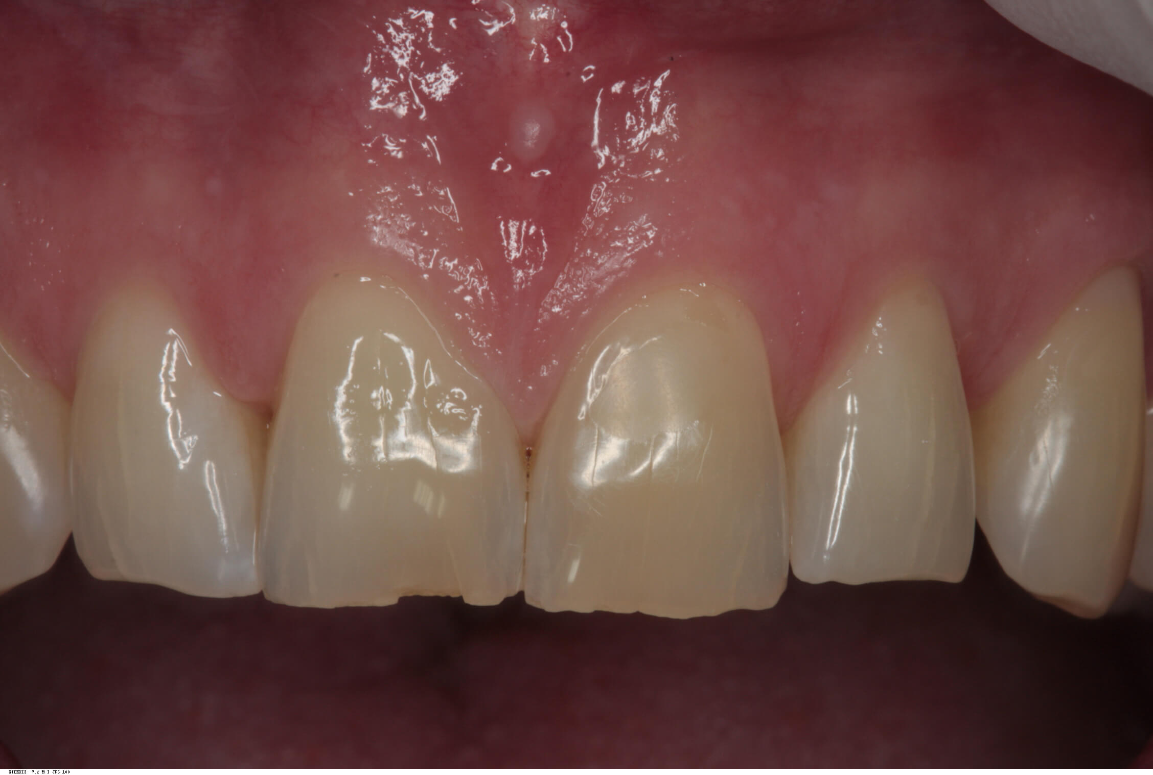
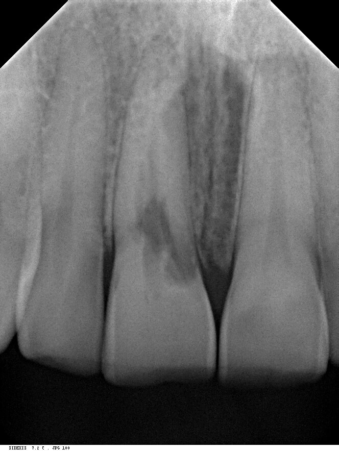
The radiograph shows extensive internal root resorption making this tooth completely unrestorable. It also shows excellent bone height into proximally both at the mesial and the distal.
Post Extraction
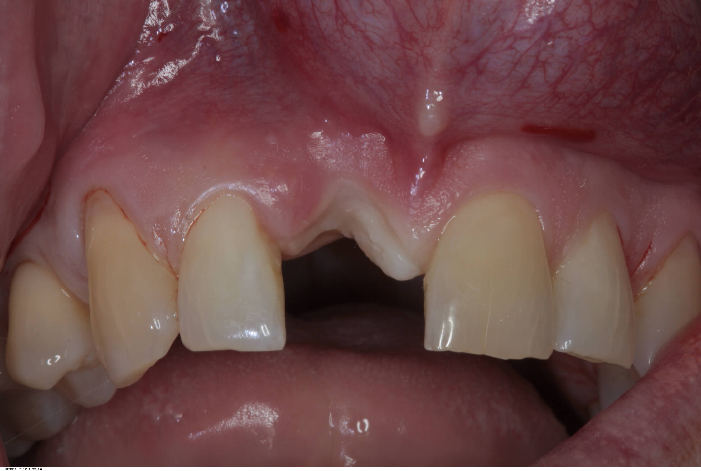
Due to the patient’s thin biotype, and very little existing labial plate seen on the cone beam radiograph, this treatment was carried out as a delayed two stage procedure. Therefore, the tooth was extracted and a provisional partial denture provided until complete soft tissue healing had occurred eight weeks later. This follows the early placement protocol as described in the ITI study guides.
Treatment
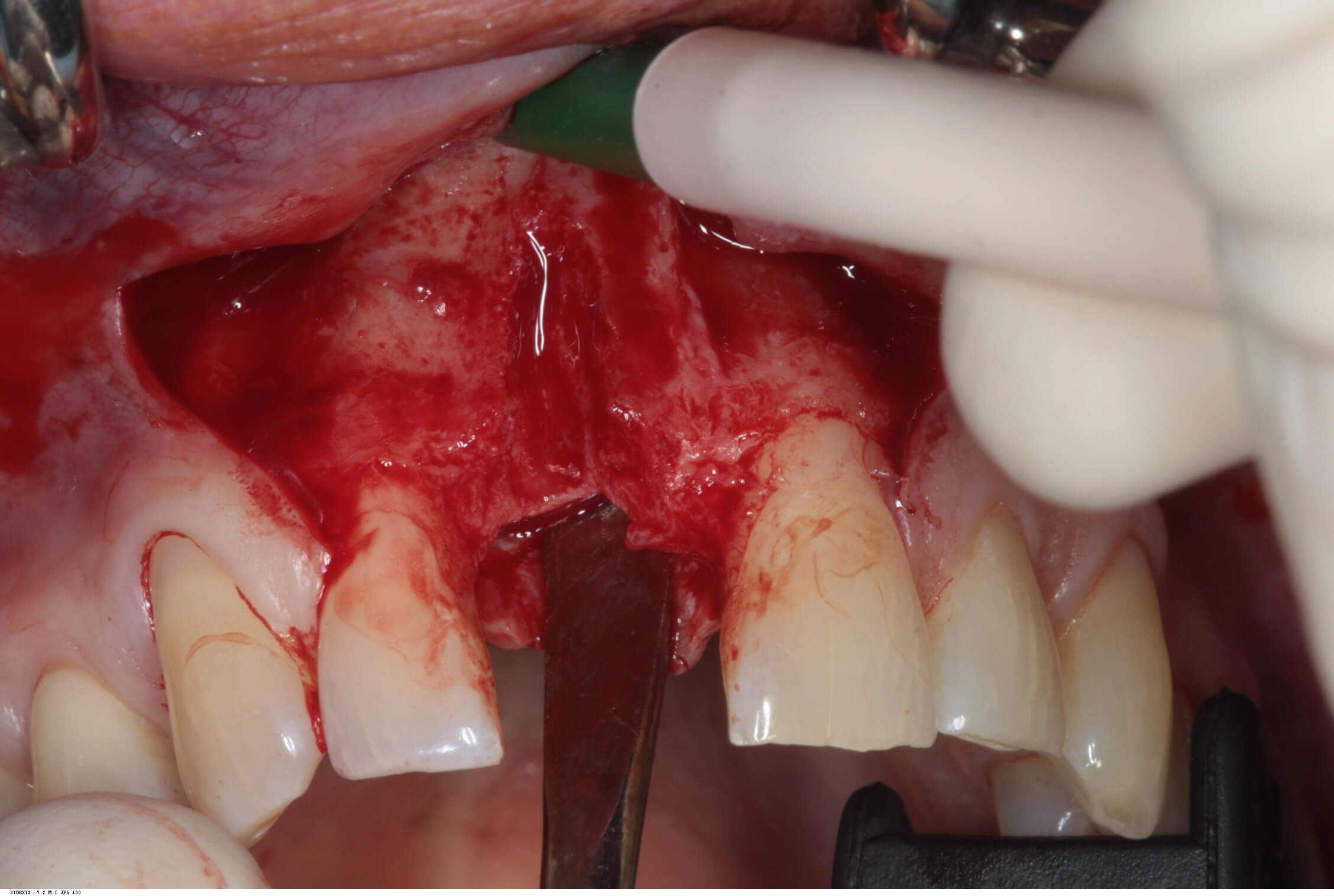
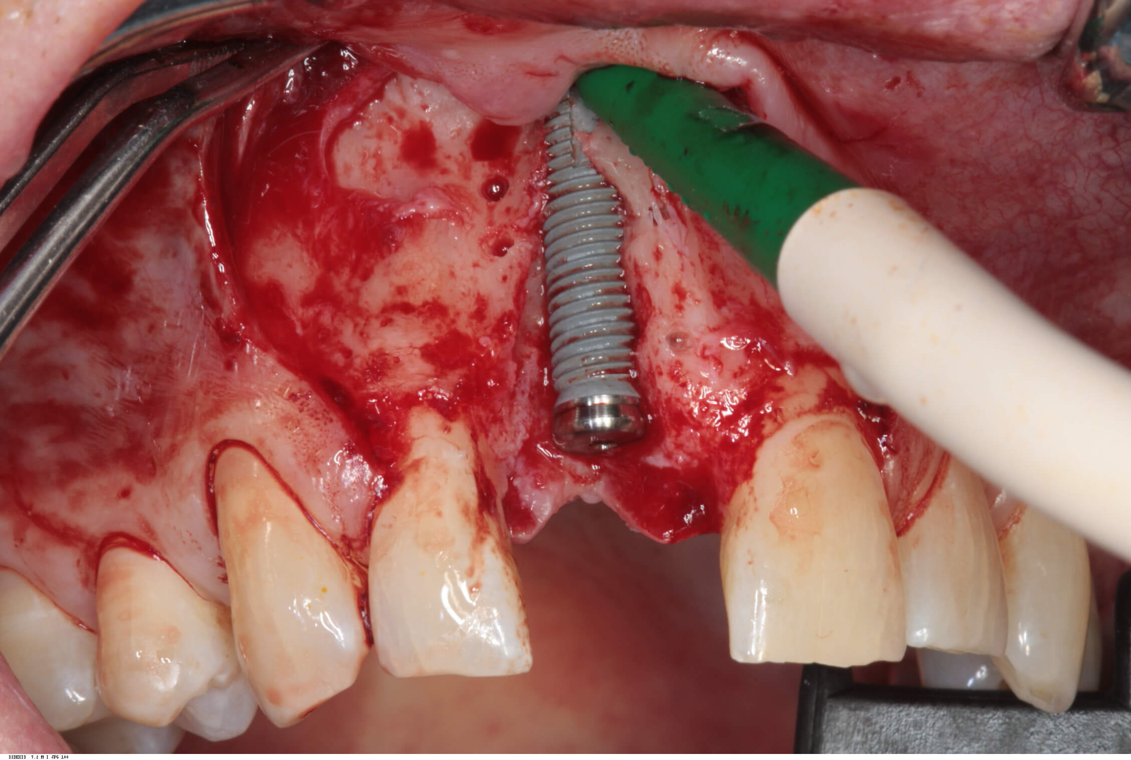
A full thickness flap was raised extending from the distal of 12 to the distal of 21. A Branemark Mark IV implant was placed achieving good primary stability, and the labial plate in the vicinity of the implant perforated to increase blood supply to the planned bone graft.
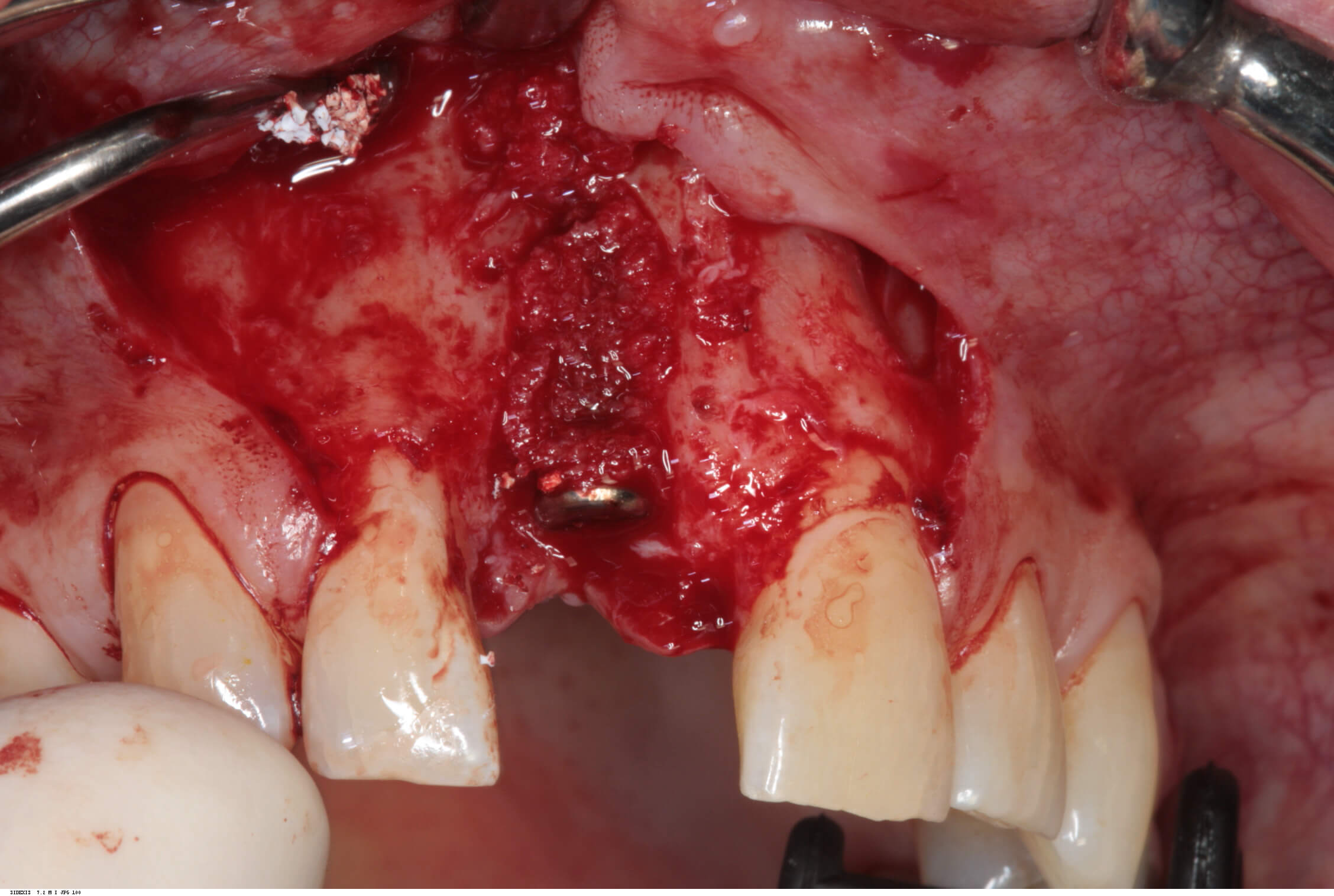
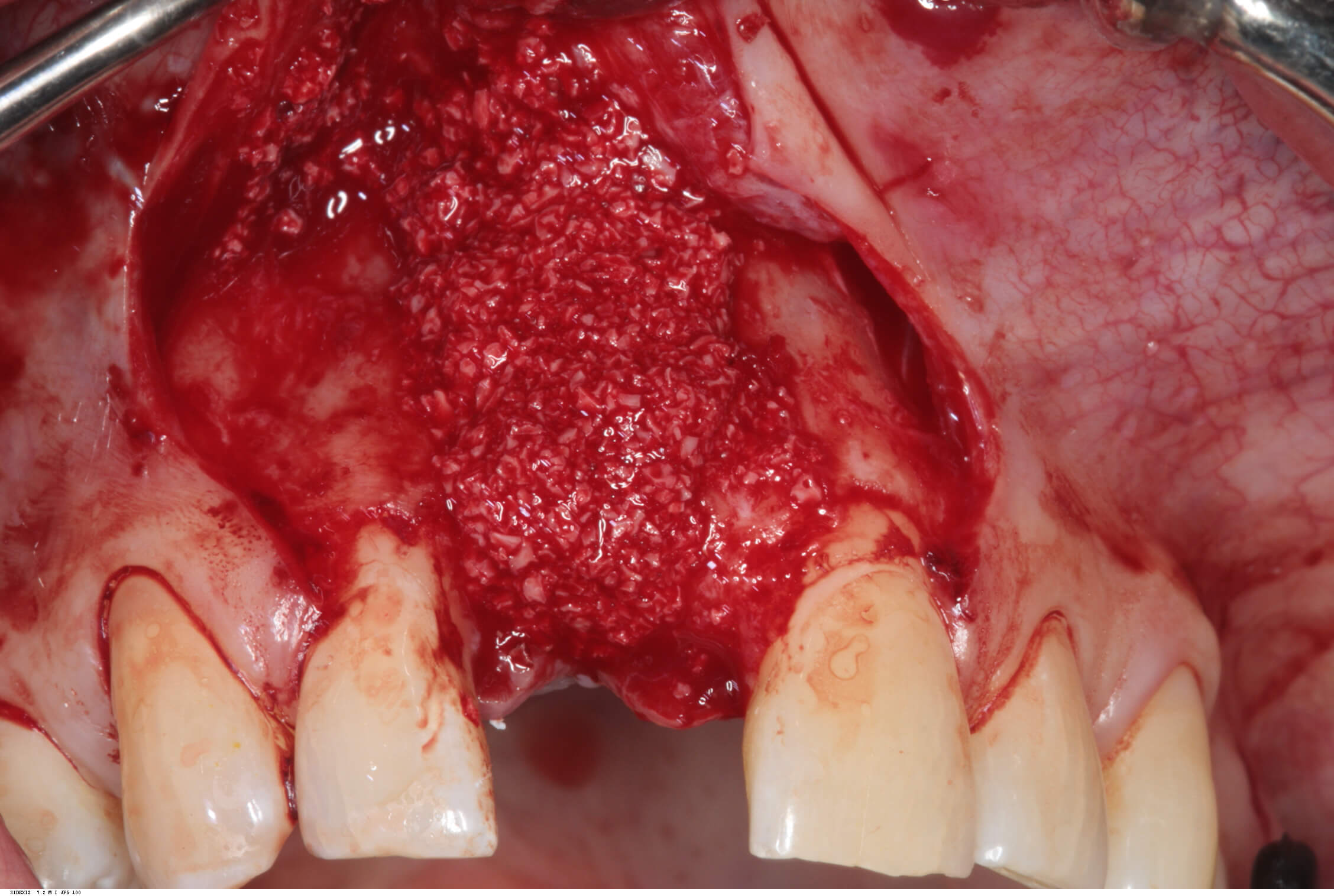
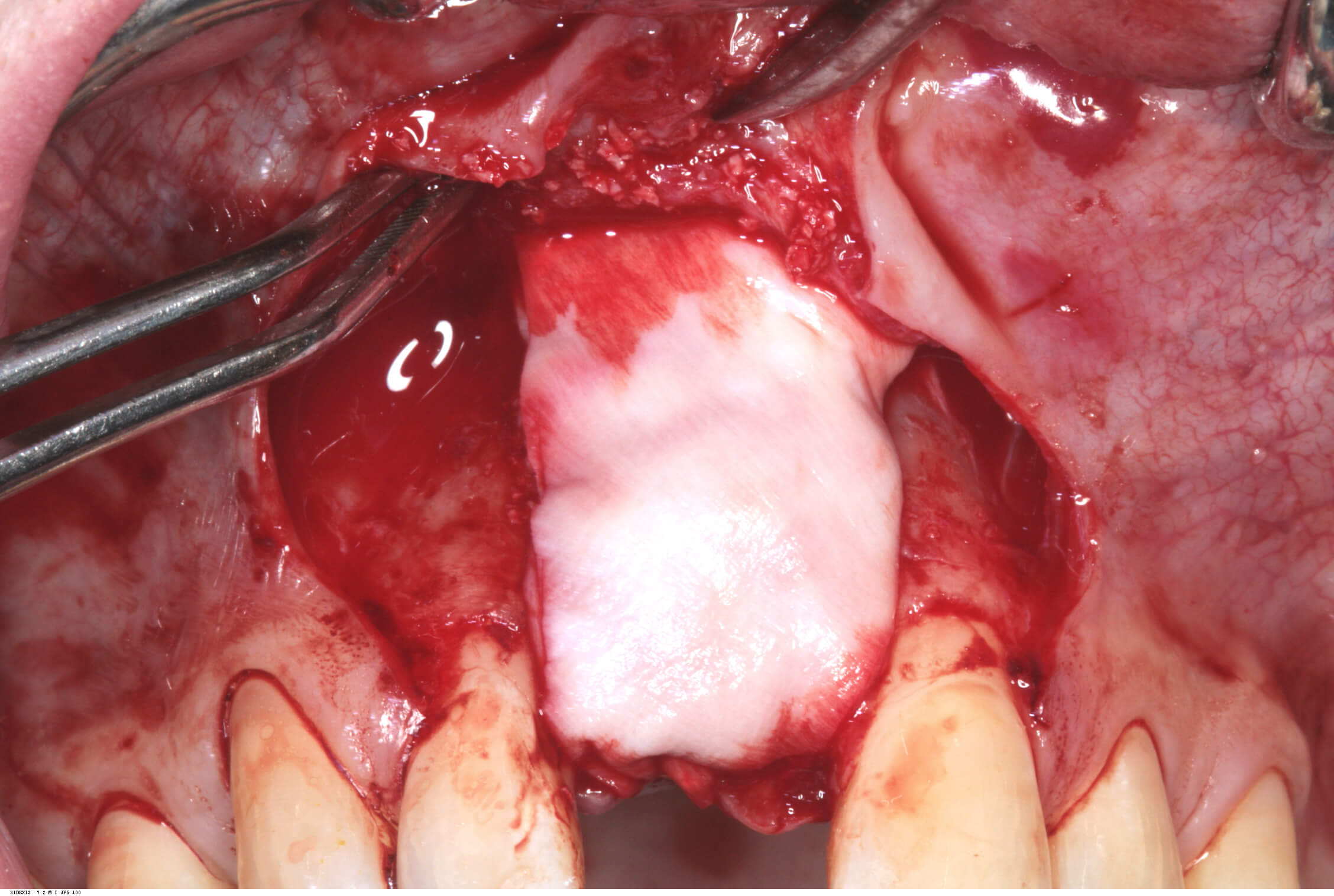
Autogenous bone chips were then harvested from the local area and placed over the exposed implant threads. This was then protected by a layer of Bioss previously mixed with blood, and then two layers of Bioguide.
The flap was then extended slightly at the relieving incisions, and releasing incisions were made to ensure a completely tension free primary closure. Two mattress sutures (4/0 Vicryl) were used along with multiple interrupted (5/0 Vicryl) sutures.
Following a four month post-operative review and confirmation of integration, this implant was then restored with a screw retained integrated abutment / veneered zirconia crown. The crown was then inserted and abutment screw torqued to 35 Ncm. The access cavity was restored with PTFe tape and composite resin. The occlusion was adjusted to ensure light Shim stock hold in ICP, and smooth concave protrusive guidance shared with the neighbouring central incisor.
Review
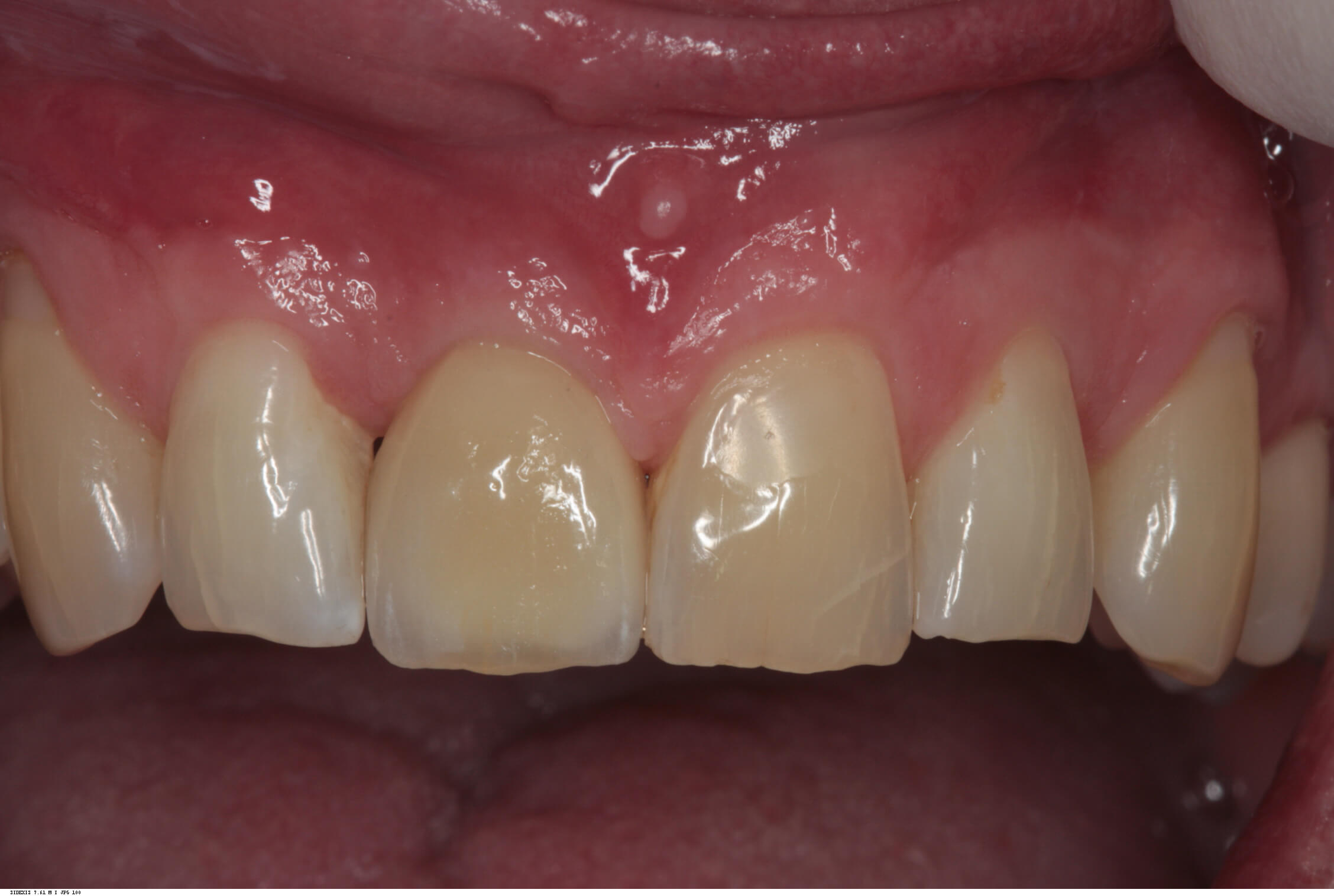
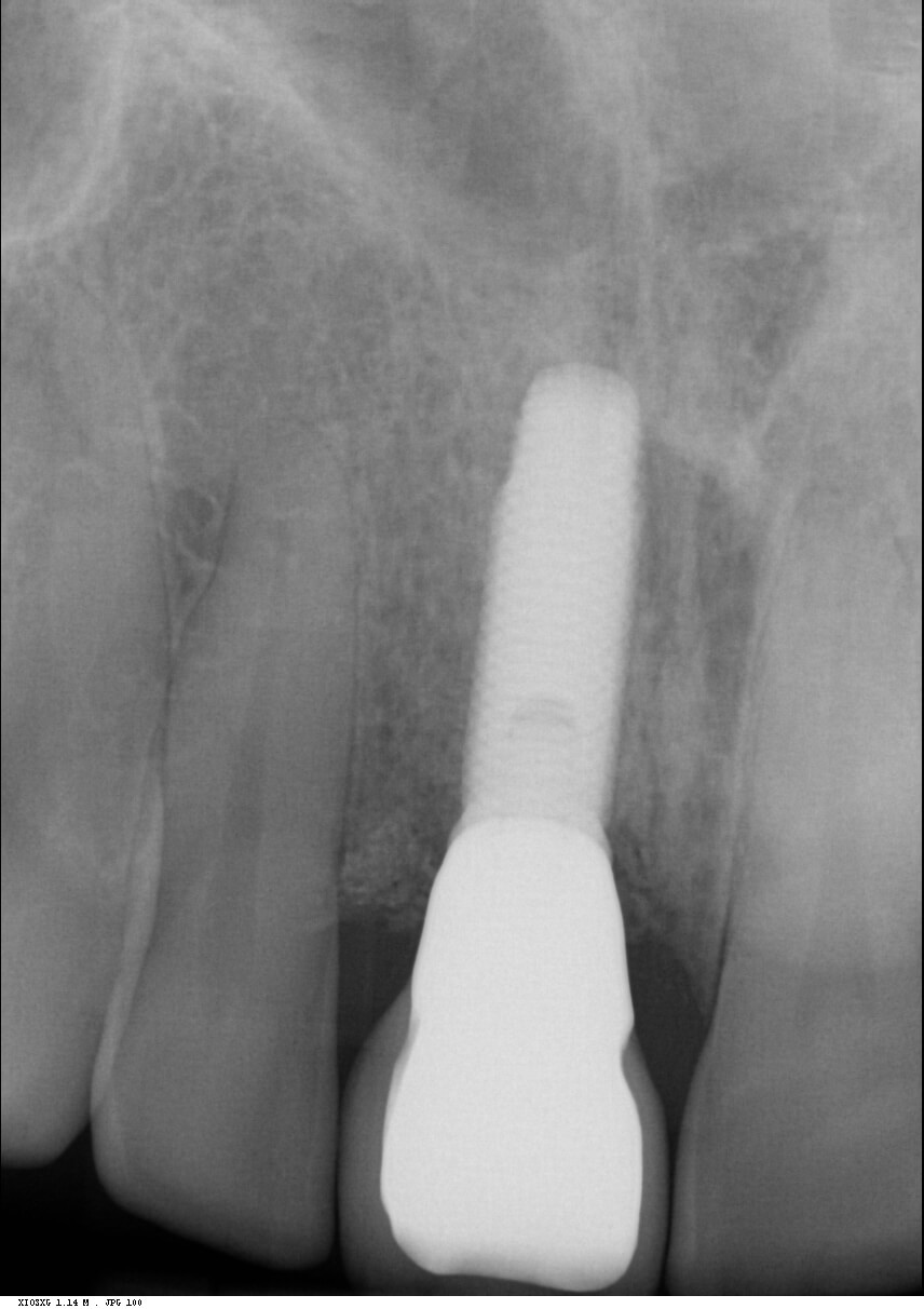

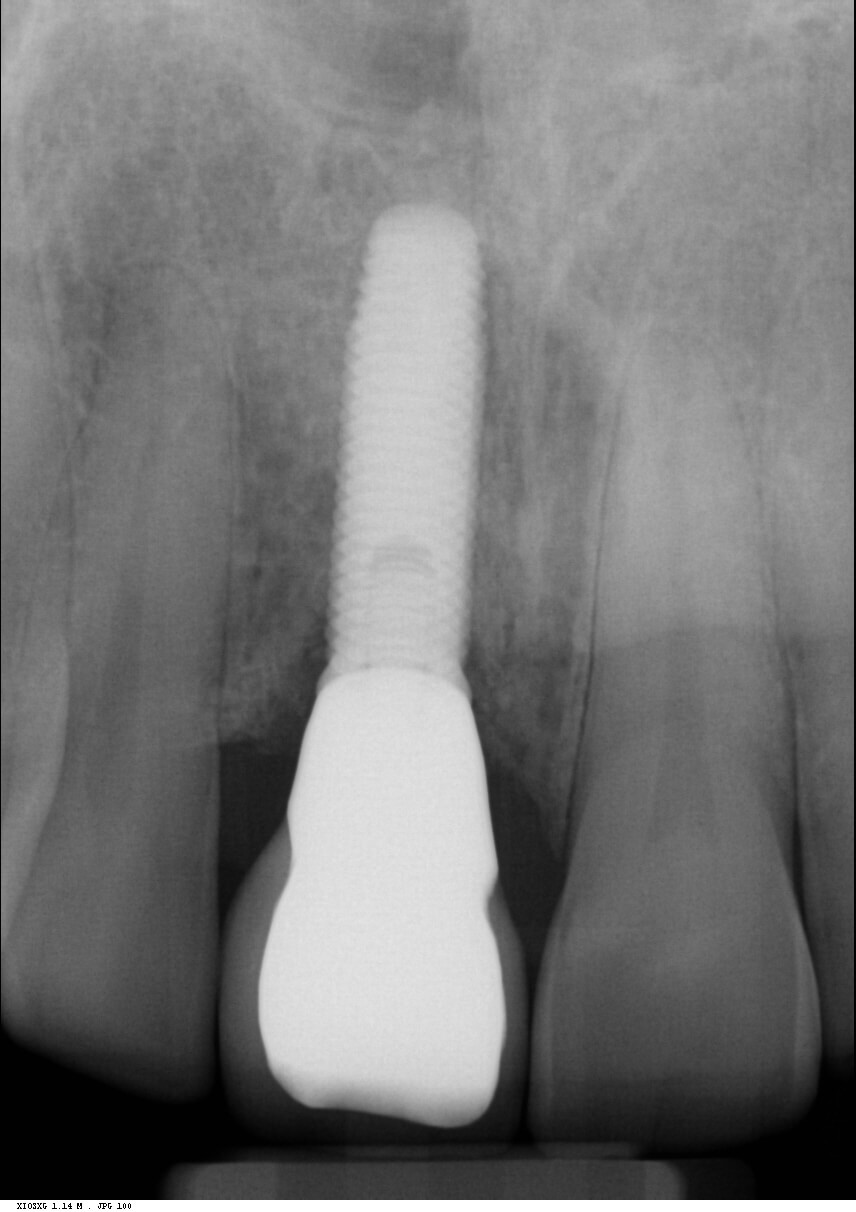
Reviews carried out at both one year and then six years post operative, showed stable gingival symmetry, and the radiographs showed excellent bone level maintenance.