Complete Maxillary Implant-Supported Bridge Case
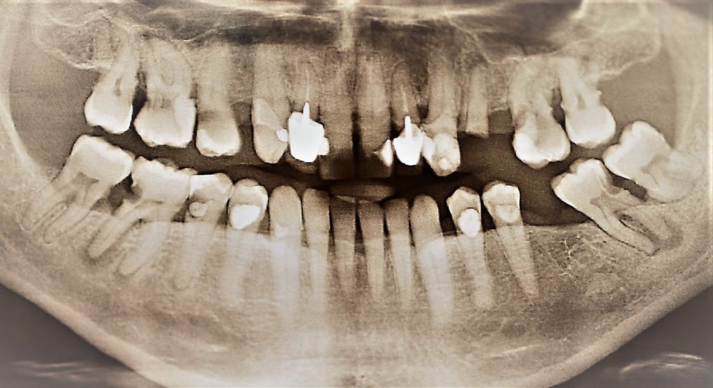
A healthy 45-year-old male patient presented with multiple large carious lesions in his maxilla and posterior mandible. Following careful treatment planning discussion along with all options, we agreed to carry out full maxillary clearance along with removal of his posterior mandibular molars. We would then place six Bioconcept tissue level implants as a one stage surgical procedure under minor oral sedation, and restore with a complete porcelain fused to zirconia, 12-unit screw-retained bridge.
Restoration of non vital tooth 45 would be carried out by his referring general dental practitioner.
Extractions were carried out as a staged approach to allow for provision of an immediate complete maxillary denture. Posterior units were extracted and allowed to heal for eight weeks before complete clearance and immediate provision of the complete maxillary denture.
This allowed for ideal Establishment of aesthetics, phonetics and occlusion in the removable prosthesis prior to provision of the definitive implant supported restoration.
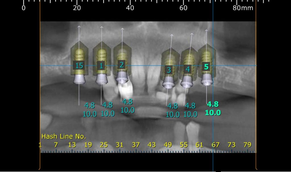
A CBCT was taken to assess bone volume available for implant placement, along with possible need for grafting, and ideal positioning. 3D planning software was used, along with a surgical guide, fabricated from a copy of the complete maxillary provisional denture.
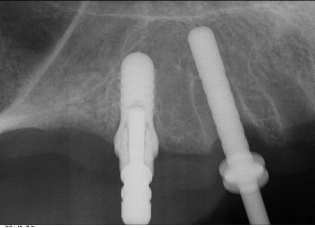
Multiple intra-operative radiographs were taken using depth gauges of gradually increasing diameter to ensure ideal positioning, particularly near vital structures such as the maxillary sinus.
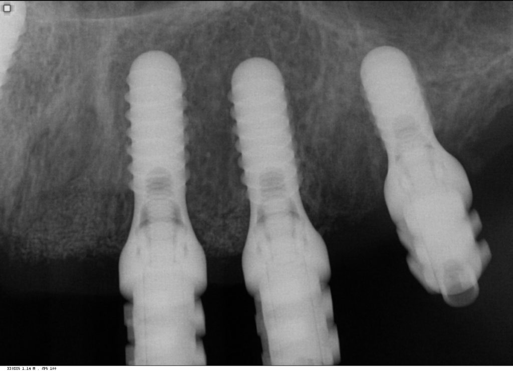
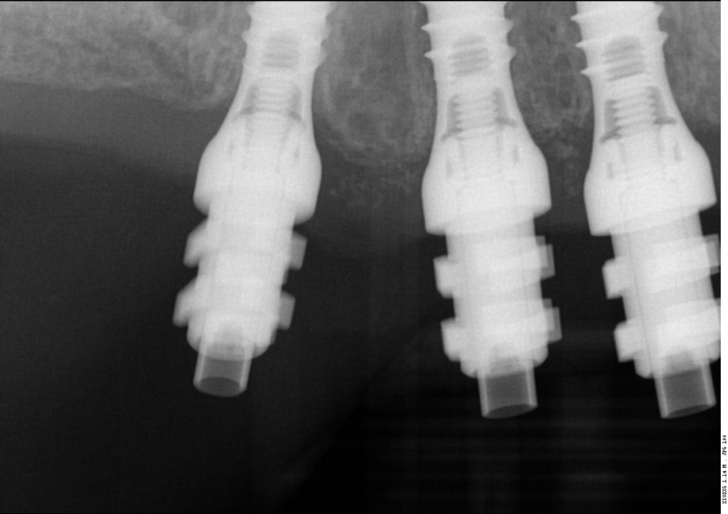
Following three months healing, definitive impressions were taken. Radiographs were taken to confirm complete seating of the impression copings prior to splinting of the impression copings and then impression taking.
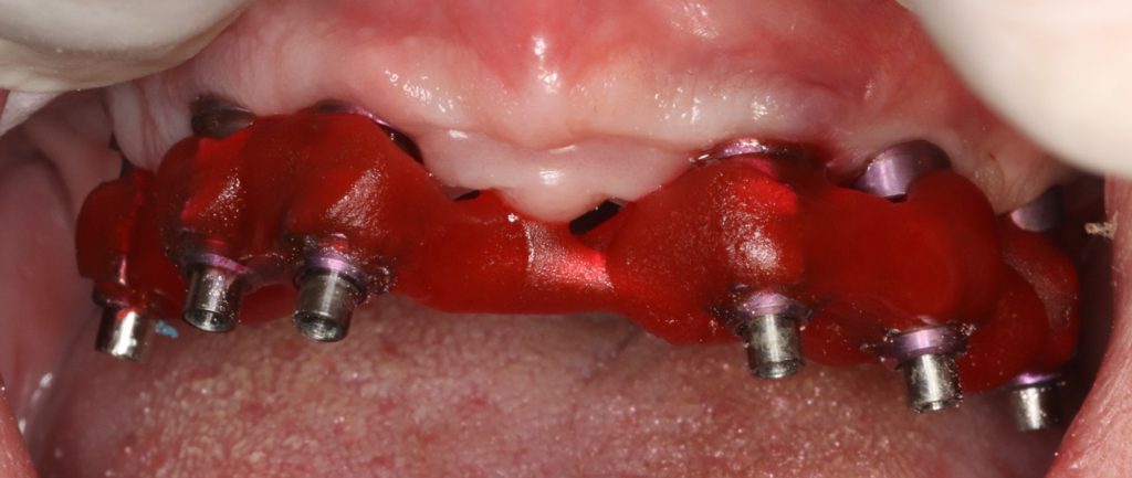
Impression copings were splinted together using metal wire and gradual incremental buildup with Fuji Pattern resin (GC Corp). Care was taken to avoid buildup of stresses in the resin leading to distortion in the impression.

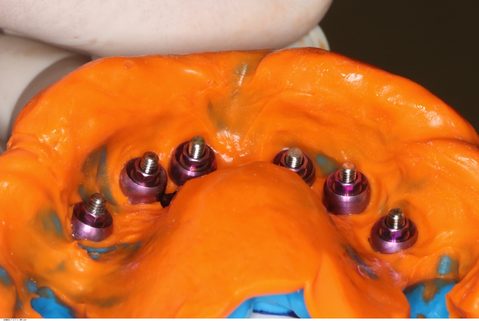
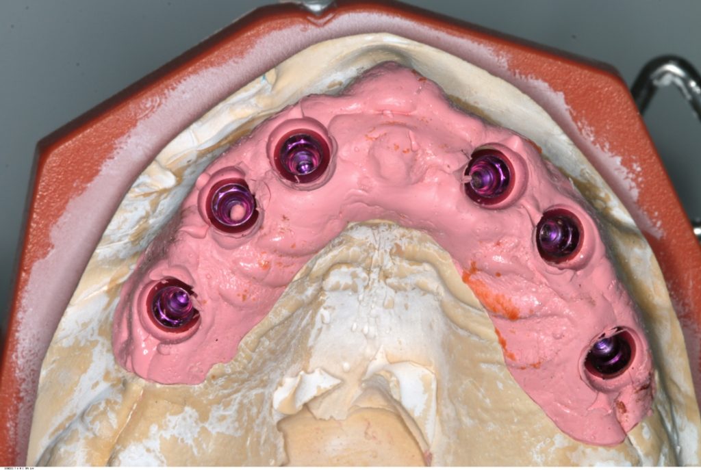
Addition-cured PVS impression material (Take One, Kerr) was allowed to complete the set before removal. Silicon-based trays are often used so that the guide pins are easily accessible prior to removal. impressions were poured and master cast were constructed in the laboratory
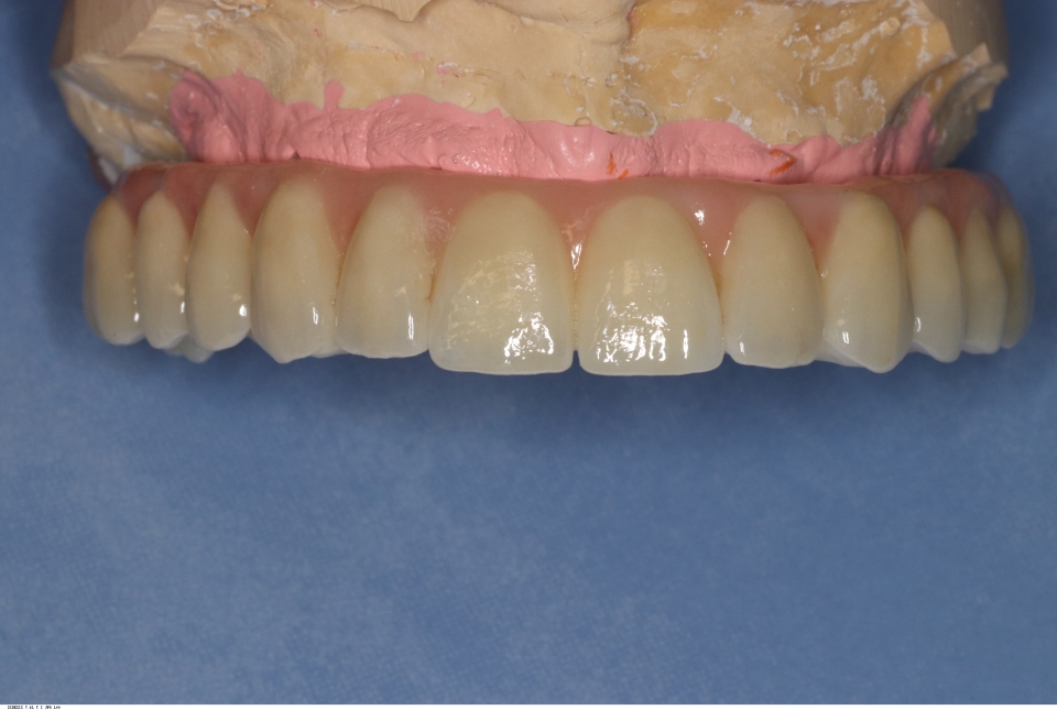
Records stage: due to the fact that all records were gained by the adjustment and confirmation of the provisional removable complete maxillary denture, records were quite straightforward. The complete denture was copied, flanges were removed, and the resulting guide was then seated over the existing tissue level healing abutments. A PMMA bridge with plastic inserts was constructed digitally in the laboratory and sent for final confirmation prior to construction of the definitive porcelain-fused-to-zirconia bridge.
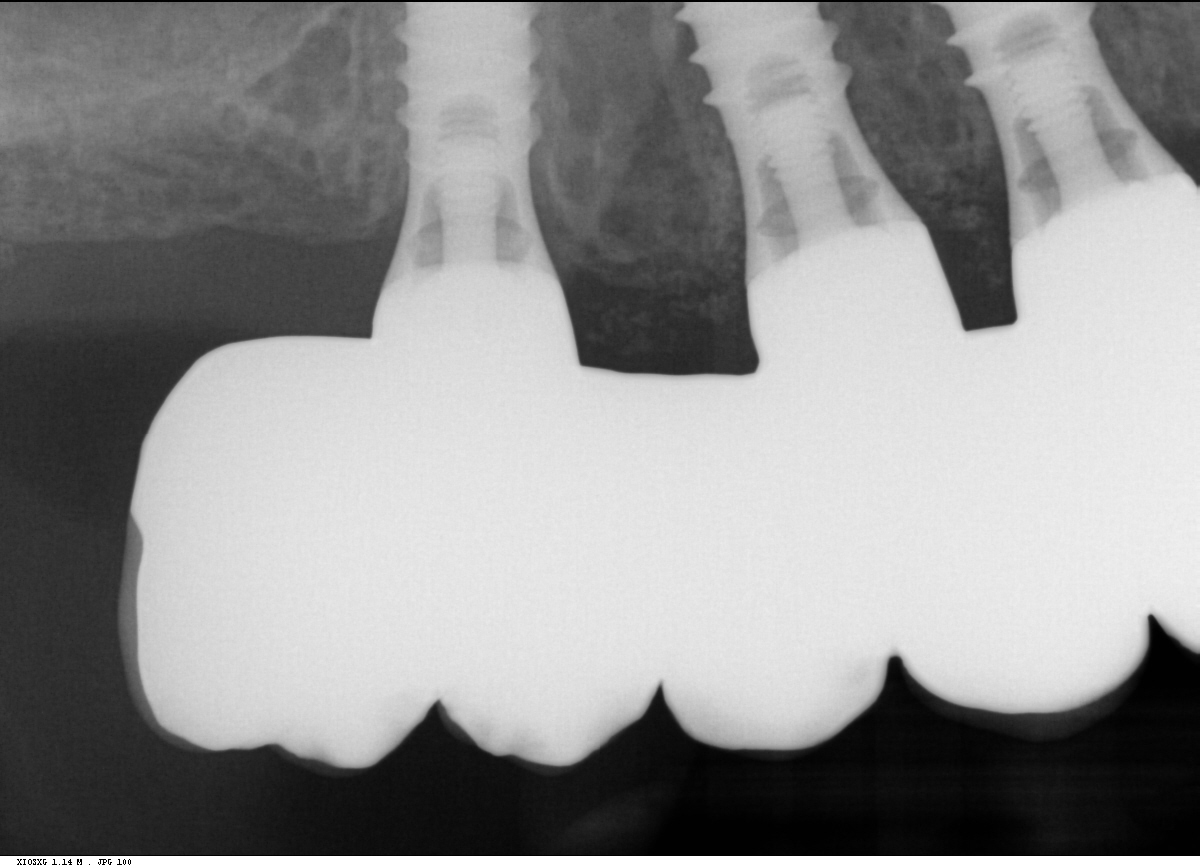
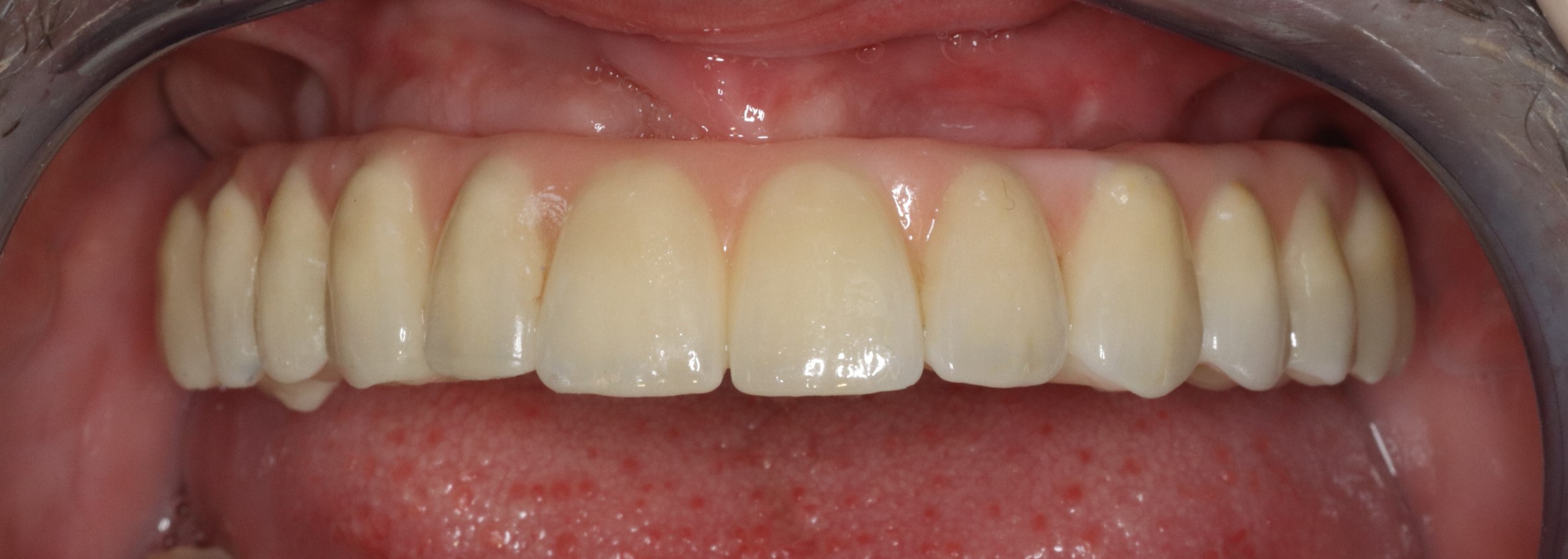
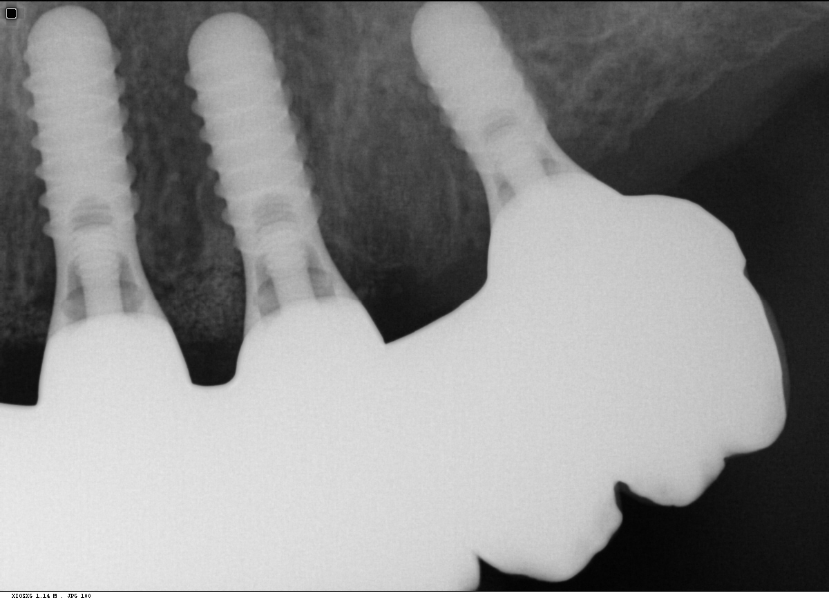
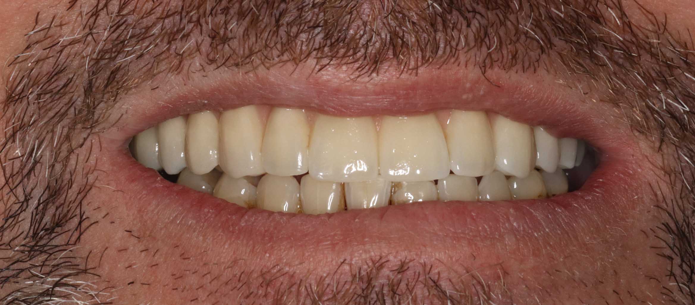
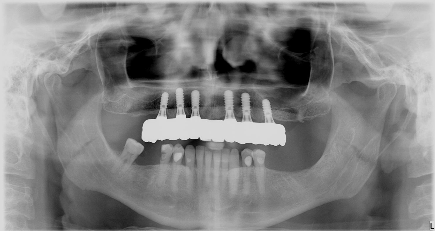
The definitive bridge was inserted and radiographs were taken to confirm full seating and correct emergence profile. Once passivity of fit was confirmed, abutments crews were torque to 35 Ncm, and the access cavities were restored with PTFe tape and composite resin. The occlusion was adjusted to ensure guiding services were smooth, concave, and gradually increasing pre-truce in steepness, and smooth shared group function in lateral excursion.
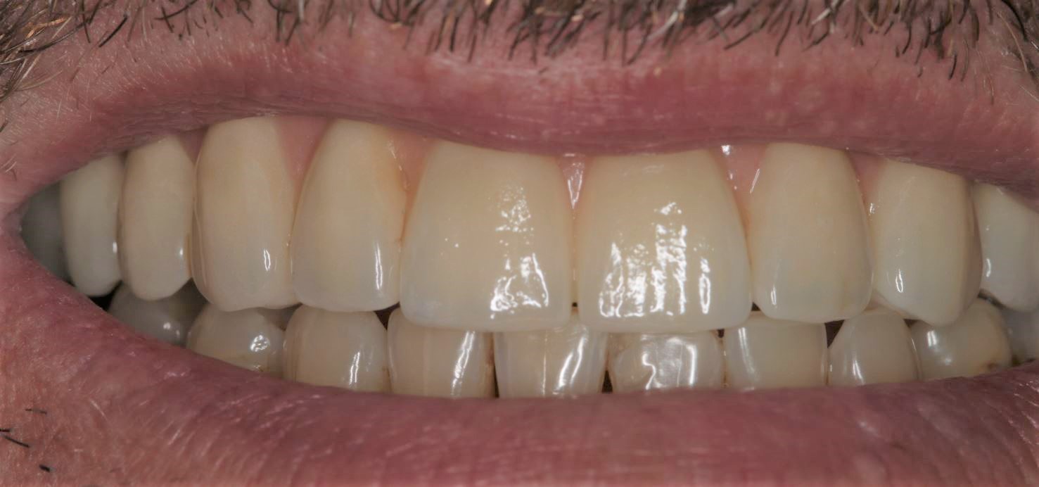
Photo and X-ray at 2 year review