Bridge in the Posterior Mandible
This healthy 71 year old female patient presented with an unrestorable right mandibular posterior four unit bridge, due to catastrophic fracture of the mesial retaining tooth 44.
Diagnostic Pre-Treatment Images
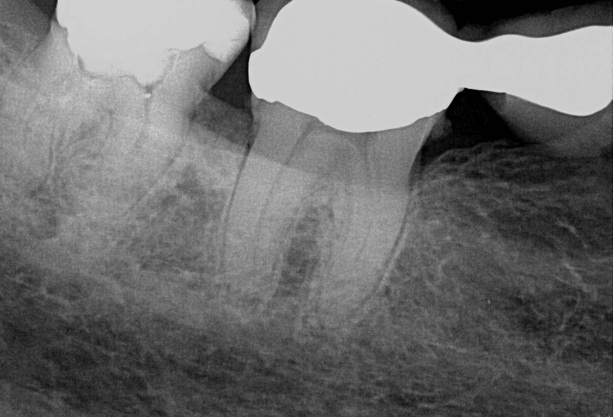
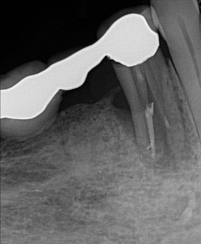
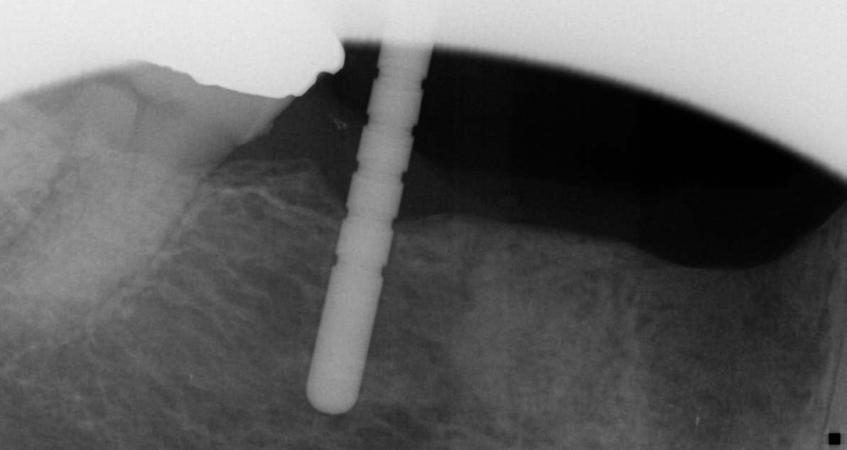
Due to the extent of the infection, tooth 44 was extracted, the site carefully curetted, and then left to heal for three months prior to bone volume assessment with a cone beam tomograph (Soridex).
Treatment Images
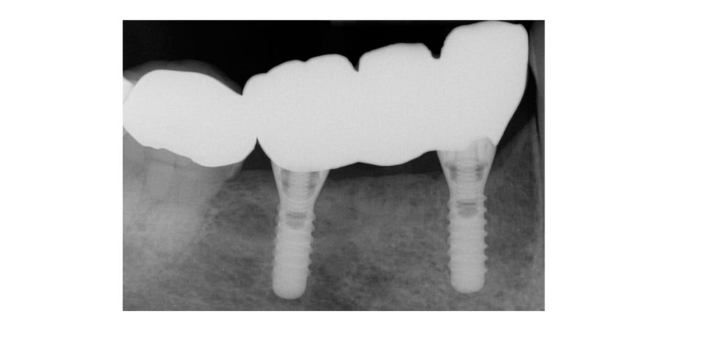
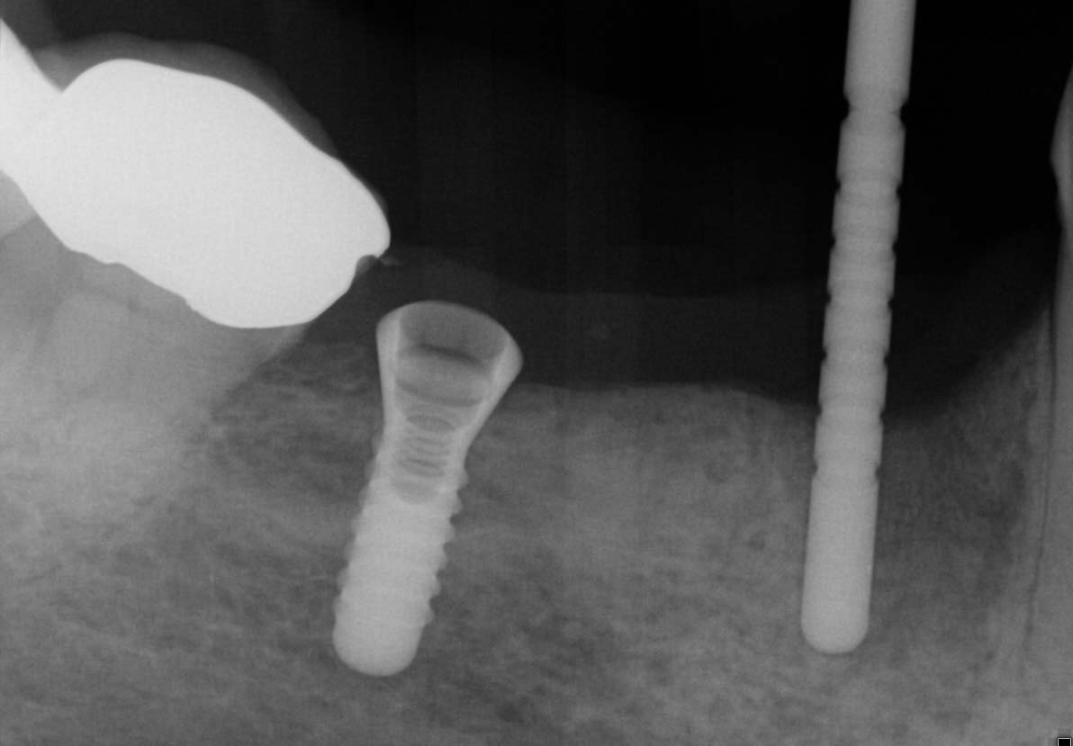
Once healing and adequate bone volume above the mandibular canal had been confirmed, two Bioconcept 3.3mm x 10mm tissue level implants were placed in sites 44 and 46. Several careful progressive intraoperative periapical radiographs were taken to confirm vertical position relative to the mandibular canal. Both implants were then placed with a primary stability of over 30 Ncm as a one stage surgical procedure, and 2mm healing abutments were then secured.
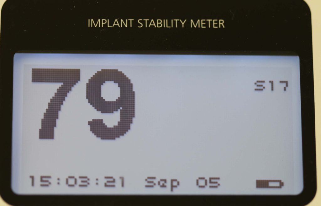
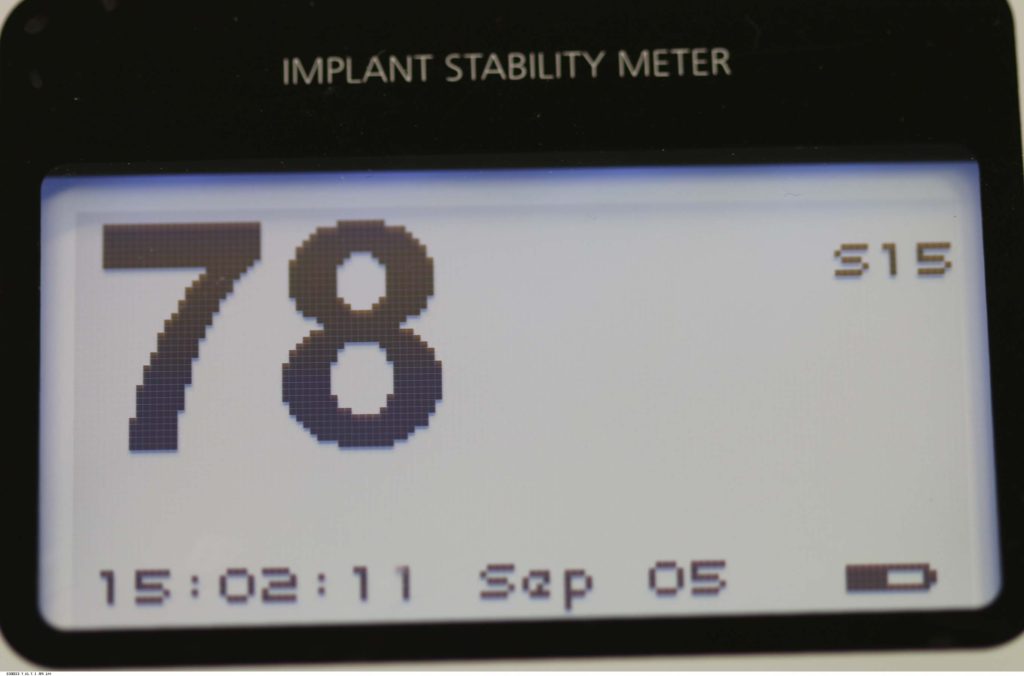
Following three months healing time, osseo-integration was then confirmed with the usual methods, combined with ISQ measurements (Ostell), to confirm readings of over 65 for adequate stability to allow for restoration.
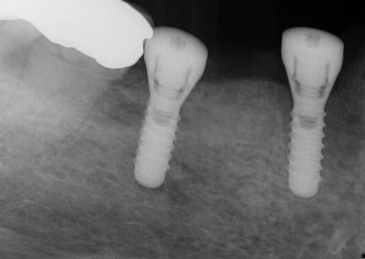
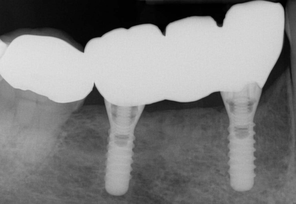
Open tray screw retained impression copings were then attached and confirmed with radiographs, and a three-unit veneered zirconia bridge was constructed using Variobase lab cemented abutments to provide an all-in-one screw-retained restoration.
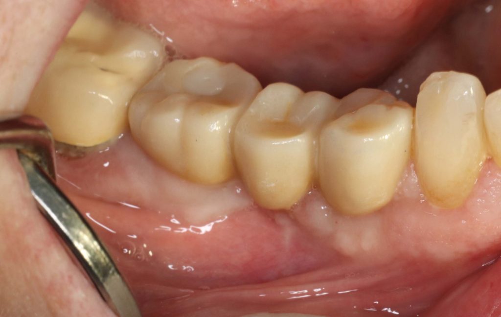
The definitive restoration was then inserted and checked for passivity of fit. The abutment screws were then definitively torqued to 35 Ncm. The access cavities were restored with PTFE tape and composite resin.